Tuesday, April 30, 2013

Pediatric Nephrology

Pediatric Nephrology


1. Renal Tubular Acidosis
• Hyperchloremic, Non-Anion Gap, Metabolic acidosis
– Alkali loss: Intestine or Kidney?
• What should the kidney do in Acidosis?
– Secrete acid and retain alkali.
• Acid urine (U pH < 5.5)
• If the kidney is not excreting acid and retaining HCO3 then something is amiss?


Proximal RTA (type 2)
– Proximal Tubule
– HCO3 re-absorption is Dysfunctional 
.
Proximal RTA

Impaired proximal tubule (PT) HCO3 absorption
• HCO3
reabsorption in PT is decreased
• Resulting in “Extra HCO3” in the urine (FeHCO3>5%)
• Serum HCO3 decreased
•Urine pH 
• Low in acidosis
• High with treatment


• Distal RTA (type 1 & 4)
– Distal Tubule
– H+ secretion is Dysfunctional 


Distal RTA type 1
Impaired Distal Tubule acid secretion
• Hydrogen ion secretion in the Distal Tubule is impaired.
• K+ reabsorption is impaired.
• H+ is not available to convert NH3 to NH4
+
• Limited acid excretion and new HCO3 creation.
• Urine pH is always high (>5.5)


Urine pH in RTA - not useful in diagnosing RTA
Can be High or Low
• Proximal RTA
– When Plasma HCO3 is low, the impaired proximal tubule can handle the reabsorption load resulting in an:
– Acidic urine.
– When plasma HCO3 is normalized (with treatment), the impaired proximal tubule cannot handle the filtered load resulting in an:
– Alkaline Urine.
• Distal RTA
– Final acidification of the urine occurs in the distal tubule via H+ secretion. This is defective in Distal RTA and hence:
– Urine pH is always aAkaline


Urine Anion Gap: 
Urine (Na+ + K+) - Cl-
• Useful to differentiate the type of RTA
– It tells us about NH4
+ excretion:
U AG = Na+ + K+ + NH4 + = Cl- + HCO3
-
• Urine AG is Negative in Proximal RTA or healthy kidney
• Urine AG is Positive in Distal RTA.
• U AG is not accurate if:
– Dehydration, unmeasured ions are present (ketoacidosis) or 
diuretics are in use.


How to Diagnose RTA?
• RTA likely exists when you have:
– Hyperchloremic, non-anion gap, metabolic acidosis.
– No extra-renal (i.e. GI) HCO3 losses
– No excessive intake of a Cl- containing acid
• Urine pH and Urine anion gap are useful to 
differentiate the type of RTA.


Why do we care about RTA?
• Chronic acidosis leads to: 
– Polyuria, failure to thrive and growth delay.
• Chronic hypokalemia causes: 
– Nephrogenic D.I., urinary retention, ileus, muscle weakness / rhabdomyolysis and rarely cardiac arryhmias.
• Calciuria in Distal RTA leads to: 
– Rickets and Kidney Stones.
Associations with structural, auto-immune or metabolic disease:
– Obstructive Uropathy
– Cystinosis, Glycogen storage disease, Lowe’s syndrome, DM I & II
– Sjogren syndrome, Hypoaldosteronism
– Etc.


Management of RTA
1. Oral alkali: 1- 5 meq/kg/day*
– Na-Bicarbonate
– K- Citrate
*Alkali needs lessen with age
2. Evaluate and treat associated conditions.


Classification of RTA
• Proximal RTA (type 2)
– Dysfunction of proximal tubule re-absorption of filtered HCO3 .
– Often seen with generalized proximal tubular dysfunction 
Fanconi Syndrome: wasting of K+, HCO3, AAs, glucose, LMW protein, etc.
– Clinical Association: Cystinosis and Lowe’s Syndrome.
• Distal RTA (type 1 & 4)
– Dysfunction of distal H+ secretion and Ammonium (NH4+) generation
– 2 types: 
• Type 1 - Low K+
• Type 4 - High K+ (due to lack of aldosterone activity)
Clinical Association: Obstructive Uropathy, Hypoaldosteronism, Drugs or Chronic renal failure.


Metabolic Alkalosis and Hypokalemia

  • Tubular Disorders
    • Bartter’s Syndrome
      • Defect in the Furosemide -sensitive distal Na-K-2Cl Transporter:
      • • Salt (NaCl) wasting => polyuria, dehydration=> Renin-angiotensinaldosterone activation => Hypokalemic Alkalosis.
      • • Birth Hx: Polyhydramnios
      • • S/S: 
        • – Dehydration, constipation, vomiting, muscle weakness.
        • – Polydipsia, polyuria, salt craving
        • – Urine Calcium wasting
        • – Failure to thrive and short stature
        • – Hyper-Reninemia, but NO HTN
    • Gitelman Syndrome
      • Similar to Bartter’s, but milder.
      • Hypokalemic Metabolic Alkalosis.
      • Low –Magneseum level.
      • Defect in the Thiazide sensitive Na-Cl transporter.
      • High Renin, but no hypertension. (increase in vasodilatory PGs)
      • May present later in life with fewer symptoms
    • Liddle Syndrome
      • hypokalemic alkalosis plus HTN
      • Primary increase in collecting duct Na+ reabsorption and K+ secretion.
  • Hyper-Aldosteronism -

    • hypokalemic alkalosis plus HTN
  • Persistent Vomiting 
    • hypokalemic alkalosis


  • Self-Induced Metabolic Alkalosis
    • • Surreptitious Vomiting or Diuretics
    • – Anorexia / Bulemia
    • • Dental erosions, calluses or ulcers on dorsum of hand, puffy cheeks (salivary gland hypertrophy).
  • – Urine Cl-
    • • Low with chronic vomiting or prior diuretics
    • • High with diuretics or Bartter / Gitelman
    • – Check Urine diuretic levels if needed.


  • Diuretic abuse


Monday, April 29, 2013

Chronic Kidney Disease

 Chronic Kidney Disease


Creatine levels difficult to interpret

example:
At What Level of Creatinine Does a 65-Year-Old Diabetic, Hypertensive White Woman Weighing 50 Kilograms Have CKD?
• 77% said:
Creatinine > 1.5 mg / dl
– GFR = 37 mL/min/ 1.73 m2
– Ccreat = 30 mL/min
BUT
• Creatinine = 1.0 for GFR = 59 mL/min/1.73 m2


The Risk of Kidney Failure is Not Uniform
Relative risks compared to Whites:
African Americans 3.8 X
Native Americans 2.0 X
Asians/Pacific Islander 1.3 X
The relative risk of Hispanics compared to non-Hispanics is about 1.5 X

Risk factors for CKD
Nonmodifiable risk factor  
  • Older age 
  • Black Race 
  • Genotype 
  • Prematurity 


Modifiable risk factor

  • Hypertension
  • Proteinuria
  • Dyslipidemia
  • Hyperuricemia
  • Smoking


Mechanisms of Kidney Disease Progression
• Adaptive changes lead to maladaptive consequences
• Hypertension
• Hyperfiltration
– Elevated glomerular pressure
• Glomerular growth
– Increased wall stress
• Increased ammoniagenesis
• Complement activation and tubulo-interstitial disease

There are ~12 million adults in the US with CKD 3 or greater, but only ~500,000-600,000 patients with ESRD or a kidney transplant.
Why the disparity????

Far more CKD patients die due to CVD than reach ESRD
• CKD is considered by some to be a coronary equivalent
– Data indicate that patients with lower GFRs (<45ml/min) and microalbuminuria or proteinuria carry a very high CVD risk (~ equivalent to a prior history of coronary disease).

Risk Factors for CVD in CKD
• Age
• Diabetes mellitus
• Smoking
• Hypertension
• Dyslipidemia
• Physical inactivity
• Menopause
• Obesity
• Anemia
• Hyperparathyroidism
Hyperphosphatemia*
• Hypocalcemia
• Effects of dialysis 
• Hypoalbuminemia and malnutrition
• Systemic inflammation
• Hyperhomocysteinemia
• Volume overload


UREMIA = urine in the blood

Uremia as a Clinical Syndrome
• Renal excretory failure
• Retained products of metabolism
• Related to protein intake
• Partially dialyzable
• Exact nature is unknown
• E.g. Small molecules (Urea etc.), lipid soluble molecules, middle molecules – urea (BUN), hormones, polyamines, middle molecules, serum proteases, trace elements, pyridine derivatives, 2-microglobulin
• Loss of metabolic and endocrine functions normally performed by the intact kidney

Uremia: Common Symptoms
• GI Nausea, vomiting, diarrhea
• CVS Dyspnea, edema, chest pain
• Neuro Restless legs, twitching, confusion
• Skin Pruritus, bruising, uremic frost
• MSK Bone pain, arthritis

Uremia: The Common Signs
• Sallow pallor, bruising
• Uremic fetor
• Hypertension
• Pericardial rub
• Alteration of consciousness
• Neuropathy

Alonzo Morning _NBA star FSGS

Adaptation of kidney function


Mechanisms of Adaptive Natriuresis in CKD
• Signal: ECF volume expansion
• Potential effectors
– Atrial natriuretic peptide (ANP)
– Other circulating natriuretic factors
– Local renal vasoactive factors

Clinical Manifestation of Sodium Balance in CKD 
• Common  ("Hoagie Night")
– Weight gain
– Peripheral edema
– Pulmonary edema
• Uncommon
– Renal Na wasting (ECF volume depletion)
– Weight loss
– Systemic hypotension

Potassium Homeostasis

Renal K Handling in CKD
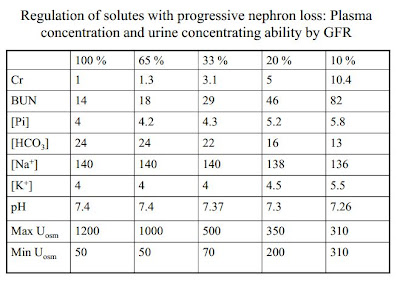
  • • K balance is very well maintained with progressive nephron loss
  • • Filtered load of K decreases as GFR falls
  • • K reabsorption is similar in normal and diseased kidneys
  • • Adaptation: Distal K secretion is increased in proportion to the decrement in GFR
    • via aldosterone
Mechanisms of Adaptive K Secretion in CKD
• Enhanced Na,K-ATPase activity
– Extracellular K concentration
– Aldosterone
• Increased distal tubular flow
– Adaptive natriuresis
– Osmotic diuresis per nephron
– Chronic metabolic acidosis

Hyperkalemia in CKD
• Aldosterone-related
– Hypoaldosteronism
• Idiopathic (Diabetes Mellitus)
• ACE inhibitors
– Pseudohypoaldosteronism
• K-sparing diuretics
• Distal flow-related
– ECF volume depletion
– Congestive heart failure (without diuretics)
• Insulin-related
– Diabetes mellitus
– Fasting
– Malnutrition
• Miscellaneous
– B-adrenergic agonists (esp. in diabetics)
– Dietary indiscretion

Anemia and Kidney Failure
Primary factors
  • Relative erythropoietin deficiency
  • Shortened rbc lifespan
    • “uremic toxins”
    • EPO may be a red blood cell “survival factor”
  • Inhibitors of erythropoiesis--”uremic toxins”
Features of the anemia of CKD:
  • Normocytic and normochromic
  • Low reticulocyte count
  • Normal bone marrow--and usually not needed to diagnose
  • Serum erythropoieitin level low-normal--not needed to diagnose

Treatment of CKD Anemia
  • Give EPO?
  • but normalizing Hb leads to higher rate of death in CKD patients
  • cut off is Hb or 7-8 g/dL

*Phosphate Homeostasis

coronary calcifications occur with high PO4 x Ca

Phosphate Homeostasis review articel


Bone/Ca/Phos/PTH Management
  • • Low Phosphate diet
  • • Maintain Ca X Phos product less than 55mg2/dl2
  • • Phosphate binders with meals 
    • positive charge binders; e.g., calcium (Tums with meals)
  • Use of Calcitriol or D3 analogues
    • dampens stimulation of PTH
  • • Monitor Ca, Phos, PTH
  • • Avoid the use of aluminum
    • neurotoxic


Thursday, April 25, 2013

Acid Base 1

Acid Base 1

Kidney can acidify urine down to a pH of about 4.1.  This is not acid enough to get rid of the daily fixed acid load of about 80 mEq/day.  Therefore buffers are essential for survival.  Phosphate buffer is limited due to low plasma concentration.  Ammonium buffer is unlimited due to unlimited supply of glutamine.

Contraction alkalosis


K homeostasis


Handy Tools

Respiratory acidosis or alkalosis = rule of 12

acute:  if PCO2 is changed by 12, the pH should change by 0.1
e.g., PCO2 = 52, pH = 7.3
        PCO2 = 28, pH = 7.5

if pH does not follow this rule, there is compensation (needs days to happen)

Metabolic acidosis or alkalosis = rule of 6

acute:  if HCO3 is changed by 6, the pH should change by 0.1
e.g., HCO3 = 18, pH = 7.3
        HCO3 = 30, pH = 7.5

if pH does not follow this rule, there is compensation (needs seconds to happen)

Wednesday, April 24, 2013

Acid Base 2

Acid Base

Anion Gap = measured cations - measured anions
in practice: AG = Na+ - (HCO3- + Cl-)

Metabolic Acidosis
AG metabolic acidosis  =  dec. HCO3- with nl chloride and unmeasured anion; e.g., lactate

"make it "

•Uremia: failure to excrete endogenous organic acids
• Ketoacidosis: metabolism of fatty acids in diabetics or alcoholics
• Lactate: tissue hypoxia from sepsis, CO or cyanide poisoning, cardiac arrest, etc

"or take it"

• Ethanol
• Methanol
• Ethylene glycol
• Salicylates
• Paraldehyde


Traditional mnemonic:
– E- ethanol
– M- methanol
– U- uremia
– D- diabetic ketoacidosis
– P- paraldehyde
– I- Inhalants (CO in particular)
– L- Lactate
– E- Ethylene glycol
– S- Salicylates

Non AG metabolic acidosis = dec. HCO3- with inc. Cl-; e.g., aceylzolamide inhibition of carbonic anhydrase in proximal tubule


• GI tract
– Diarrhea
– Pancreatic fistula
– Ureteral diversion into ileal loop (bladder removal with ureter put into ileum)


• Kidney
– Renal tubular acidosis
– Carbonic anhydrase inhibitor (acetazolamide/Diamox)


Osmolar Gap  = calculated - measured osmolarity
Measurement of the serum osmolal gap is most useful in patients with a high anion gap metabolic acidosis, particularly when methanol or ethylene glycol poisoning is suspected

"make it"

Ketoacids
Uremia

"or take it"

Drug metabolites
Methanol, Ethanol
Other alcohols

Metabolic Alkalosis

vomiting = H+ loss.  and inc. plasma HCO3
volume depletion make alkalosis worse due to RAAS system - low urine chloride

contraction alkalosis - vomiting


Hyperaldosteronism


Tuesday, April 23, 2013

Acute kidney injury

Dr. Xu - acute kidney injury


GFR

Each kidney contains approximately 1 million nephrons. 
GFR is the sum total of the filtering rates of all 2 million nephrons in the kidneys.
Single nephron GFR x # of functioning nephrons.
Normal value for GFR depends on age, sex, and body size, and is approximately
130 mL/min/1.73 m2
for men and and 120 mL/min/1.73 m2
for women.
How much plasma does the kidneys filter in a day?
• 130 ml/min x 1440 min = 187 liters or roughly 180 liters per day.
• Average plasma volume is about 3 liters. 
• 180 L/ 3 L= 60. Plasma gets filtered 60x in a day!

Pathophysiokogy of AKI

In hypovolemia, as RPF falls, kidney compensates to maintain a stable glomerular filtration pressure by 2 mechanisms. 
1. Vasodilation of the afferent arteriole via local prostaglandin production.
2. Efferent vasoconstriction via angiotensin II. 


Volume depletion from any etiology will decrease renal perfusion and renal plasma flow (RPF). 

2. Decrease in RPF, if uncompensated by several autoregulatory mechanisms, will decrease GFR. 
3. Result is prerenal AKI.

Prerenal AKI

kidney normal but perfusion low


BUN/Cr > 20
Normal BUN: Cr ratio is < 10. BUN:Cr ratio > 20 in prerenal AKI.
• Increased BUN reabsorption in prerenal AKI is caused by the increase in 
the passive reabsorption of urea that follows the increased proximal 
sodium reabsorption.


FeNa < 1%  (avid Na reabsorption)  falsely high with diuretics

Situations Where FENA < 1% Does not Indicate Prerenal AKI
• Contrast nephropathy
• Rhabdomyolysis
• Acute GN
• Hepatorenal syndrome
• ATN with CHF
• ATN with cirrhosis
• ATN with severe burns

FeUrea < 35%


Typical Features of Prerenal AKI
• Clinical history pointing to hypovolemia is most important clue.
• BUN/Cr ratio > 20:1. Normal BUN/Cr ratio is 10. 
• UNa < 20 mEq/L
• FENa < 1%
• FEUrea < 35% (Useful if patient on diuretics) 
• Urine sediment: bland or hyaline casts


Treatment of Prerenal AKI
• Pre-renal AKI is not pathology. It is physiology. 
• Normally functioning kidneys are simply responding to volume depletion with end result being Cr rise. 
• IVF replacement is treatment in patients who are volume depleted.

Intrarenal AKI

oliguric (urine output < 500 ml/24 hrs)


Most damage in ATN is to proximal tubule (where 70% of Na is normally reabsorbed)

Mechanism of Oliguria in ATN – Tubuloglomerular Feedback
• 70% of ATN cases will cause oliguria (UOP < 500 ml/day).
• Glomeruli are actually intact in ATN. So why are they not filtering enough plasma 
to generate urine?
• Total body water (TBW) accounts for about 50% of body weight. So an 80 kg person has 40 L of TBW. 1/3 of this 40 L is ECF, so that’s 13 L. 1/3 of ECF is intravascular fluid. So the plasma volume is 13 L x 1/3 = 4L.
• Healthy kidneys can filter 100 ml/min of plasma. That means it takes about 4000/100 = 40 min to filter all the plasma in the body. 
• 9% of the filtered plasma is reabsorbed by the tubules and returned to circulation. 
• In ATN, tubular reabsorption is impaired. If the glomeruli keep on filtering while the tubules are not reabsorbing, then a person can become volume depleted.
• So oliguria is really an adaptive response in ATN.  "Acute Renal Success"


Mechanism of Oliguria in ATN – Tubuloglomerular Feedback
• How does GFR get turned down in ATN?
• Tubuloglomerular feedback.
• ATN with tubular injury  reduced NaCl reabsorption at the PCT  increase NaCl delivery to macula densa at DCT  chemoreceptor activated and releases vasoactive compounds  afferent arteriolar vasoconstriction and a fall in GFR  less filtration  which in turn limits any further NaCl loss.


Treatment in ATN
• Supportive care only. IVF to keep MAP > 65 mmHg to ensure renal perfusion.
• Lasix does not make ATN better.
• Lasix can be used to control volume overload. Worth a try.
• When does a patient with ATN need dialysis? % fluid overload > 10% (about 8 kg 
fluid up)
• Indications for acute hemodialysis:
1. Acidosis (refractory pH < 7.1)
2. Electrolyte (refractory K+ > 6.5, hypercalcemia > 13 with oliguria)
3. Intoxication (ethylene glycol, salicylate poisonings)
4. Overload of volume (unable to maintain O2 sat) 
5. Uremia (MS change, nausea and vomiting)

Acute Interstitial Nephritis (AIN)


Etiology of AIN
• Medications account for 70% of AIN. Big offenders are:
1. Penicillins
2. Quinolones
3. Bactrim
4. PPIs
5. Diuretics
• Infections account for 10% of AIN
1. CMV
2. Legionella
3. HIV
• Rest from miscellaneous causes, such as:
1. Autoimmune disorders like SLE, Sjogren’s
2. TINU syndrome (tubulointerstitial nephritis with uveiitis)
3. Hypercalcemia (recent case at UNM)


Clinical Features of AIN
• Asymptomatic
• Constitutional symptoms (fever, chills, malaise, etc)
• Triad: Fever, Rash, Eosinophilia (5-10%)
• Flank tenderness
• Non oliguric AKI
Eosinophiluria  Neither sensitive nor specific


Treatment of AIN
• Discontinue suspected offending agent. Should see improvement within 3-7 days.
• If Cr does not improve 3-7 days after dc offending agent, consider biopsy and or a trial of steroid at 1 mg/kg up to 60 mg daily for one month with fast taper
• Patients with drug-related AIN should improve within 1-2 weeks after steroid therapy.
• Complete recovery may take up to 6 weeks.

Glomerular Nephritis


Clinical Features of Glomerulonephritis
Nephrotic syndrome:
1. Massive proteinuria > 3 g/day
2. Fluid overload: HTN, edema
3. Due to noninflammatory glomerular injury
Nephritic syndrome:
1. Mild proteinuria < 3 g/day
2. Prominent hematuria
3. Acanthocyte (dysmorphic RBC) on urine microscopy
4. RBC casts on urine microscopy
5. Due to inflammatory glomerular injury

Postrenal AKI


Pathogenesis of Postobstructive Nephropathy
1. How urine obstruction causes AKI is intuitive: renal function falls when urine flow is blocked. 
2. Actual mechanism has to do with elevated pressure transmitting back into the glomeruli.
3. Important to keep in mind that only bilateral obstruction or unilateral obstruction of a solitary functioning kidney will cause renal failure.
4. Is renal failure from obstruction reversible?
• Maybe not completely if > 2 weeks. In animal models, obstruction > 2 weeks cause interstitial fibrosis. If prolonged, this is irreversible.


Dr. Alas  -  Calcium Phosphorus  Magnesium


Plasma calcium is tightly controlled
– Total calcium 8.5-10.5 mg /dl
• Free (ionized) floating Ca is 45% of total Ca
- Approximately 4.5mg/dl-5.3 mg/dl
• Bound (reversibly) to plasma protein is 40% of total Ca
– 90% of bound Ca is to albumin
• Complexed to anions makes up 15% of total Ca
– Anions complexed to include (chloride, citrate, phosphate)
– Ca in this form is diffusable and available for filtration at the glomerulus


Ca homeostasis
• Increases serum calcium:
– Immediate release of Ca from skeleton is mediated by parathyroid hormone (PTH) acting indirectly on osteoclasts
– PTH is released to quickly restore a falling plasma calcium
– PTH also activates the vitamin D pre-hormone to increase Ca absorption from the intestine
• Decreases serum calcium:
– Calcitonin 
• C cells of the thyroid
• Inhibits osteoclast bone resorption


(Dys)Functions of Calcium
• Necessary ion for metabolic processes
– Regulate ion channels
– Promote activation of enzymes
– Structural involvement (bone and teeth)
• Neuromuscular excitability
– Tetany and spasms
– Cardiac arrhythmias
– Lethargy (hypercalcemia)(Dys)Functions of Calcium(cont’d)
• Involved in alveolar surfactants
• Coagulation
– Prothrombin  thrombin
– Fibrinogen  fibrin


Renal Handling of Ca++

Proximal Tubule★
• Sodium is actively reabsorbed in the PT
• Calcium reabsorption occurs through passive diffusion
• Ca moves along the paracellular route in the PT!!!!


Thick Ascending Loop of Henle★
• Active reabsorption of NaCl and recycling of K in luminal membrane generates a lumen 
positive potential difference (PD)
• This positive PD drives passive paracellular diffusion.
• Furosemide inhibits the Na-K-2Cl cotransporter leading to hypercalciuria. 

Calcium Sensing Receptor


★CaSR & increased Ca intake★
• Suppression of PTH
– Decrease distal tubular calcium reabsorption
• Ca binds to CaSR => arachidonic acid metabolite => inhibits the luminal K channel and the basolateral NaK-ATPase
• K recycling halts the Na-K 2Cl as well thereby stopping paracellular reabsorption of Ca


★Distal Convoluted Tubule★
• In this segment Ca transport is an active process.
• Luminal transport initially is through TRPV5 channel then bound in cytosol to calbindin-D28K protein
• Finally it exits on the basolateral side through Ca/H or Na/Ca active exchangers
• PTH, vitamin D, and cAMP stimulate Ca absorption in the DCT.

• Thiazide diuretics cause hypocalciuria
• Thiazides inhibit the Na-Cl cotransporter; causing a natriuresis
• This ECF volume contraction causes increased Na absorption in the PT, and increased Ca 
• Amiloride causes the same effect, but on the ENaC transporter in the collecting duct.


Acute treatment of hypercalcemia★
• It is important to assess volume status.
• Severe hypercalcemia will cause a diuresis
• Steps to removal of calcium in a volume depleted state:
– Hydrate well with saline (1-2 liters)
– Once hydrated give a loop diuretic


Phosphorus


Important as energy storage, a urinary buffer for H+, constituent of bone, and signal transduction.
• Many foods contain high amounts of PO4; especially deli meats, dairy products, nuts, 
chocolate, and colas.
• Total PO4 content is 700 gm; 85% of which are in bone and teeth


Rise in Phosphorus is Associated with★★Increased Mortality★
• Normal serum range 3.0-4.5 mg/dL (Why is this important?)
• Ca x PO4 solubility product determines whether Ca and PO4 precipitate within the blood stream
• “The product”>70 places patient at risk for calciphylaxis


Brief Overview of Phosphate★Regulation
• Plasma concentration of phosphate is maintained by 1,25(OH)2D, PTH and FGF-23
• A rise in plasma PO4 stimulates PTH in 3 ways
• PO4 directly stimulates PTH synthesis
• Increased serum PO4 decreases serum Ca; stimulates the calcium sensing receptor (CaSR) on the parathyroid gland
• Increased PO4 decreases 1,25(OH) 2D decreasing it’s inhibition of PTH secretion
• Increased PO4 stimulates FGF-23 expression

Renal Handling of Phosphate


90% of PO4 is filtered by the glomerulus (10% protein bound)
• 85% of PO4 reabsorbed in proximal tubule
• FGF-23 and PTH inhibit renal tubular PO4 reabsorption (phosphatonin) 


Proximal Tubule PO4 Handling★
• Na/H2PO4 cotransporter drives reabsorption
• A maximal absorption for this transporter exists (saturable)
• PTH causes endocytosis of the Na-PO4 cotransporter; inhibiting PO4 reabsorption
• Basolateral exit is not well understood

Magnesium - 


Dr. Fisher  -  Nephritic Syndrome


IgA Nephropathy - Clinical Features (I)
• Most common glomerulonephritis worldwide
– very common GN in New Mexico!
• First described by Berger (Berger’s disease)
• Often triggered by upper respiratory infection
• Presents as nephritic syndrome with:
– hematuria and/or proteinuria


IgA Nephropathy - Pathogenesis
• Respiratory infection triggers production of antibodies
• antibodies typical of mucosal immune response are:
→ Immunoglobulin A (IgA)
→ IgA forms immune complexes with antigens in circulation
• IgA antibodies are deposited in mesangium
– incite inflammation
• In Henoch-Schönlein purpura (=systemic vasculitis):
– IgA antibodies also deposited in wall of small arteries
– incite inflammationIgA Nephropathy - Light mi


IgA Nephropathy - IF and EM
• By immunofluorescence:
– granular deposits of IgA in mesangium
– “mesangial” pattern of immunofluorescence
• By electron microscopy:
– elecron dense immune deposits in mesangium
– increased mesangial cells and matrix


Rapidly Progressive Glomerulonephritis
• Rapidly progressive glomerulonephritis is clinical term, NOT a pathologic diagnosis !!!
• Includes 3 different diseases:
post-streptococcal glomerulonephritis

• a.k.a. post-infectious GN
– 1-4 weeks after streptococcal infection
– also caused by other bacterial infections (staphylococci, others)
• Most common in children 6-10 years
– much less common in adults
• In children prognosis typically excellent (i.e. complete recovery)
– prognosis tends to be worse in adults

Pathogenesis
• Immune-mediated - patients make antibodies against bacterial antigens
• Formation of immune complexes in serum
• Immune complexes get deposited in glomerulus and incite inflammation
• Pts have antibodies to streptococcal antigens in serum 
– inc. ASO titer in serum as clinical lab test
(= antistreptolysin-O titer)

Post-streptococcal GN - IF and EM
• By immunofluorescence
– GRANULAR deposits of IgG and C3 along glomerular basement membrane
"lumpy bumpy or starry sky)
• By electron microscopy
– predominantly SUBEPITHELIAL deposits (i.e. on the epithelial side of the glomerular 
basement membrane) = HUMPS
– (smaller subendothelial and mesangial deposits may be found)


pauci-immune glomerulonephritis (=ANCA disease)


anti-glomerular basement membrane glomerulonephritis (anti-GBM GM)


Thursday, April 18, 2013

Renal Pathology

Dr. Barry  Renal Pathology


Membranoproliferatve glomerulonephritis
• Characterized histologically by alterations in the basement membrane, proliferation of 
glomerular cells, and leukocyte infiltration mainly in the mesangium
• Two types MPGN Type I and II, on the basis of distinct ultrastructural, immunofluorescence, and pathologic findings
• Type I may be primary or secondary
• (Type II: dense ribbon-like membrane deposits on EM)


MPGN Type I
• Most cases in adults are secondary to an identifiable etiology:
– Hepatitis C
– Infective endocarditis
– Lupus
– Chronic indwelling catheters
– Cryoglobulinemia
– Must exclude these before dx primary idiopathic MPGN


Cryoglobulins - MPGN
• Ig complexes precipitating in vitro when cooled below body temperature. 
• Cryoglobulinemic MPGN often appears identical to MPGN on LM, IF, and EM.
• Usually Hep C related, may be idiopathic


Diabetic Nephropathy
• Kidney disease resulting from diabetes, including effects on glomeruli, vessels and tubulo-interstitium
• A leading cause of end stage kidney disease
• Renal failure is 2nd only to MI as a cause of death in DM
• 30-40% of all diabetics develop clinical evidence of nephropathy
– 50:50 Type I:Type II in absolute terms
– (Kidney disease is more likely in Type I, but there are more Type II diabetics)
• Frequency of DN is influenced by genetics
– Native Americans, Hispanics, and African Americans with Type II DM have a greater chance of developing chronic kidney disease vs whites


Proteinuria occurs in 50% of diabetics (I&II), 
• Earliest: microalbuminuria (30-300mg/day albumin) and increased GFR are important predictors of future overt diabetic nephropathy
• Overt proteinuria, usually discovered 12-22 years after onset , mild at first, then nephrotic 
• Followed by progressive loss of GFR, leading to end stage failure within 5 years
• Without treatment, 80% of Type I and 20-40% of type II will develop overt nephropathy with macroalbuminuria, over 10-15 years
• Progression variable, but by 20 yr, >75% Type I and 20%Type II will progress to ESRD


DN arteriole hyalinosis


Pathogenesis of diabetic 
glomerulosclerosis

1. Metabolic defect (incl altered glycosylation)
– GBM thickening, increase mesangial matrix, loss 
of podocytes

• 2. Hemodynamic changes
– Similar to adaptive responses in secondary FSGS: 
increased GFR (hyperfiltration), increased 
glomerular capillar pressures, glomerular 

hypertrophy, also loss of podocytes


Summary of DN
Diabetic Nephropathy
• Thickened basement membranes
• Diffuse mesangial sclerosis
• Nodular glomerulosclerosis (KW disease)
• Global glomeruosclerosis
• Hyaline deposits
• “Capsular drops”
• “Fibrin caps”
• Hyalinizing arteriolar sclerosis
• Tubular atrophy and intersitial fibrosis

Renal Amyloidosis
Proteinuria or nephrotic syndrome
• Renal insufficiency is common

Amyloid is a pathological proteinaceous substance that results 
from abnormal folding of proteins


not a single disease-- generic term for a heterogeneous group 
of diseases that have in common tissue deposits of 
extracellular fibrillar proteins that aggerate to form a crossbeta-pleated sheets, stain positively for congo red (with 
green birefringence), and form non-branching fibrils 7.5 - 10 
nm on EM


Once deposited, amyloid encroaches upon adjacent cells and 
disrupts function


Normally light chains are reabsorbed in the proximal tubule
With a plasma cell dyscrasia  excess light chain  reabsorptive 
capacity is exceeded 


light chains in urine: “Bence Jones proteins


Light-chain cast nephropathy (myeloma kidney)
• Bence-Jones (light chain) proteinuria: the light chains may combine with Tamm-Horsfall 
protein under acidic conditions to form large distinctive casts that obstruct the tubular 
lumens and induce an inflammatory response
• Precipitated by: dehydration, hypercalcemia
• Casts: amorphous masses, fractures, may be laminated, filling/distending tubular lumens
• Inflammation: multinucleated giant cell macrophage response, granulomatous

see lecture for review slides at end