Emphysema

Normal Lung Emphysema

Normal Lung Smoker's Lung
Emphysema vs Chronic bronchitis
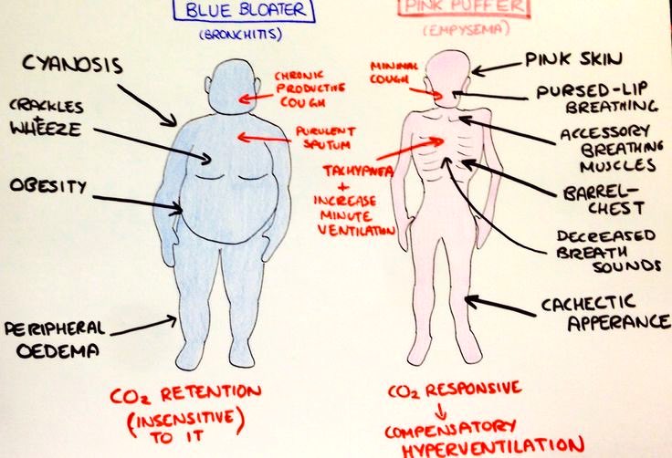
Pink puffers: emphysema
destruction of the airways distal to the terminal bronchioles--which also includes the gradual destruction of the pulmonary capillary bed and thus decreased inability to oxygenate the blood. So, not only is there less surface area for gas exchange, there is also less vascular bed for gas exchange--but less ventilation-perfusion mismatch than blue bloaters (hence: pink if compared to bronchitis). The body then has to compensate by hyperventilation (the "puffer" part). Some of the pink appearance may be due to the work (use of neck and chest muscles) these folks put into just drawing a breath.
Blue bloater: chronic bronchitis.
caused by excessive mucus production with airway obstruction resulting from hyperplasia of mucus-producing glands, goblet cell metaplasia, and chronic inflammation around bronchi. Unlike emphysema, the pulmonary capillary bed is undamaged. Instead, the body responds to the increased obstruction by decreasing ventilation and increasing cardiac output. There is severe ventilation to perfusion mismatch leading to hypoxemia and polycythemia.
Because of increasing obstruction, residual lung volume gradually increases (the "bloating" part). They are hypoxemic/cyanotic because they actually have worse hypoxemia than pink puffers
No comments:
Post a Comment