Saturday, March 18, 2017

Pulmonary Diffusing Capacity


Khan video on diffusion

This is a good video but does not mentioned the influence of capillary volume and hemoglobin.

Capillary volume is part of the surface area component (alveolar surface area + capillary volume) of the Fick Equation and is influenced by factors such as exercise which increases diffusing capacity by increasing blood flow to the top of the lungs and thereby increasing capillary volume = surface area for diffusion.  The Fick equation may be simplified by combining (A x K)/T into a single parameter, DL, the diffusing capacity of the lung.
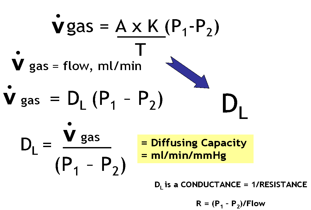


Passive diffusion proceeds at a rate proportional to the driving force (P1 – P2), surface area (A), and solubility of the diffusing gas (K) and inversely proportional to thickness of the barrier (T).  These factors comprise Fick’s law for passive diffusion, where K is Krogh’s diffusion constant. Because it is not possible to accurately measure area or thickness, these membrane properties along with K are lumped to form a parameter called diffusing capacity, DL.  As shown in the figure, the flow of gas by diffusion, in ml/min, = DL (P1 – P2).  Note that when P1 = P2, the driving pressure becomes zero and gas movement stops. This equation for gas flow can be rearranged to provide the equation for diffusing capacity:  DL = Vgas / (P1 – P2)

Hemoglobin concentration is important in the rate of diffusion because the final step in diffusion of oxygen is reaction with hemoglobin. In fact, the reaction rate of with hemoglobin accounts for about half of the total resistance to oxygen uptake in the lungs.  This means that anemia can result in impaired diffusion.
Also, as described below, diffusing capacity is measured using carbon monoxide. The binding of CO to Hb goes faster with more Hb; e.g., anemia will reduced the measured diffusing capacity.


How is Diffusing Capacity Measured?


Carbon monoxide is used to measure diffusing capacity of the lung (DL).  Advantages of CO are that its uptake is limited by membrane properties (diffusion limited) and not by blood flow (perfusion limited).  This is so because CO gas binds 100% with hemoglobin meaning there is no back pressure (P2) to slow or stop diffusion.  Since P1 is kept constant, the rate of transfer depends only on DL.

https://www.openanesthesia.org/pulm-diffusing-capacity/

Pulmonary diffusing capacity is often measured by Diffusion capacity of the Lungs for carbon monoxide (DLCO). In essence, this measures how much CO can pass from the alveoli to the blood in the pulmonary capillaries, thus giving clinicians the broader idea of how much inhaled gas can pass into the blood through the lungs. 

While some state the “DLCO correlates with the total functioning surface area of the alveolar-capillary interface (Butterworth, et al),” Dr. McCormack notes, “Older textbooks suggest that thickening of the alveolar-capillary membrane (in interstitial lung disease) and loss of alveolar membrane surface area (in emphysema) are the primary causes of a low DLCO. However, subsequent experimental data suggest these and most other diseases that influence the DLCO do so by reducing the volume of red blood cells in the pulmonary capillaries” (McCormack). Regardless of theory, whether the surface area or the alveolar surface itself is modified or the volume of the blood in the pulmonary capillaries is modified, the DLCO reflects how much gas can be transferred to the blood via the lungs.


  1. Butterworth IV, JF, Mackey DC, Wasnick JD.  Morgan & Mikhail’s Clinical Anesthesiology, 5th ed. New York, NY: McGraw Hill; 2013.
  1. McCormack, Meredith. “Diffusing Capacity for Carbon Monoxide.” Ed. James Stoller and Helen Hollingsworth. N.p., 14 Apr. 2015. Web.

Thursday, March 16, 2017

New Job in Grenada/England


Arrived in Grenada😏


View from our hotel room
SGU campus is across the harbor


Sunday, March 5, 2017


First Without Oxygen
"I am nothing more than a single narrow gasping lung, floating over the mists and summits." Reinhold Messner, Everest
Climbing Mount Everest, the tallest mountain in the world, was a challenge that eluded scores of great mountaineers until 1953, when Sir Edmund Hillary and Tenzig Norgay first reached its summit. Over the next three decades, more "firsts" followed, including the first ascent by a woman, the first solo ascent, the first traverse (up one side of the mountain and down the other) and the first descent on skis. But all of these climbers had relied on bottled oxygen to achieve their high-altitude feats. Could Mt. Everest be conquered without it?

As early as the 1920s, mountain climbers debated the pros and cons of artificial aids. One, George Leigh Mallory, argued "that the climber does best to rely on his natural abilities, which warn him whether he is overstepping the bounds of his strength. With artificial aids, he exposes himself to the possibility of sudden collapse if the apparatus fails." The philosophy that nothing should come between a climber and his mountain continued to have adherents fifty years later. 

In the 1970s, two of its strongest proponents were Reinhold Messner and Peter Habeler. Messner had achieved considerable notoriety by completing a series of spectacular Alpine rock climbs without the use of metal protection pegs. In 1974, Messner teamed up with Habeler, a quiet Mayrhofen guide who shared his philosophy, and the pair proceeded to take the climbing world by storm. Agile and slight of build, they scaled the Matterhorn and Eigerwand faces in record time. In 1975, they made a remarkable ascent of the 11th highest mountain in the world, Gasherbrum, without using supplemental oxygen. By 1978, they had set their sights on climbing Mt. Everest—without bottled oxygen.

Messner and Habeler quickly found themselves the subject of criticism by members of both the climbing and medical communities. They were labeled "lunatics," who were placing themselves at risk for severe brain damage. The physiological demands of climbing Everest had been studied on previous expeditions, and found to be extreme; in 1960-61, tests conducted on members of an expedition led by Sir Edmund Hillary concluded that oxygen levels at the summit of Mt. Everest were only enough to support a body at rest—and that the oxygen demands of a climber in motion would certainly be too great.

Despite the controversy, Messner and Habeler continued with their plan. They would climb together with the members of the Austrian Everest Expedition into the Western Cwm, and then make their own separate attempt for the summit. The teams arrived at Base Camp in March of 1978 and spent the next few weeks establishing a secure route through the Icefall, erecting camps I-V and preparing for their ascent. 

Messner and Habeler's first attempt began on April 21. They reached Camp III on the Lhotse Face on April 23. That night, Habeler became violently ill with food poisoning from a can of sardines. Messner decided to continue his ascent, without his debilitated partner, and set off with two Sherpas the next morning. Upon reaching the South Col, the three climbers were suddenly trapped in a violent storm. They battled temperatures of -40 degrees Fahrenheit and winds of 125 m.p.h. for two full days. Exhausted from struggling with a torn tent and severe hunger, even Messner later admitted to believing his venture was "impossible and senseless." Finally, a break in the weather enabled the shaken party to descend to Base Camp and recuperate.


http://www.pbs.org/wgbh/nova/everest/history/firstwoo2.html


Messner and Habeler discussed making one more bid for the summit. Habeler had begun to reconsider the use of oxygen, but Messner remained steadfast, declaring that he would not use oxygen—nor climb with anyone who was using it. He believed that climbing as high as possible, without oxygen, was more important than reaching the summit. Habeler, unable to recruit a new partner, relented, and the two became a team once more.

On May 6, Messner and Habeler set out again. They reached Camp III (7200 meters) easily and, despite a new blanket of heavy snow, felt ready to move on to the South Col the next day. They were now reaching altitudes where they could expect to feel the effects of oxygen deprivation. Messner and Habeler had agreed on carrying two oxygen cylinders to Camp IV, in case of an emergency, and had also made a pact to turn back if either person lost his coordination or speech. 

The next day, it took them only three and a half hours to reach the South Col (7986 meters), where they camped for the afternoon and evening. Habeler complained of a headache and double vision on the climb up, but felt better after resting, even though both men frequently woke up from their naps gasping for air. They forced themselves to drink tea, hoping rehydration would lessen the effect of the thin air. 

At 3 am on May 8, the two woke and began preparing for the day's attempt on the summit. Simply getting dressed took them two hours. The weather was questionable, but they decided to break camp. Since every breath was now precious, the pair began using hand signals to communicate. Progress was slow. Trekking through the deep snow was exhausting, so they were forced to climb the more challenging rock ridges. It took them four hours to reach Camp V (8500 meters), where they rested for thirty minutes. Even though the weather was still threatening, they decided to continue—at least to the South Summit, which was 260 vertical meters away. 

Messner and Habeler now faced exhaustion unlike any they'd encountered before. Every few steps, they leaned on their ice axes and gasped for breath. Messner described feeling as though he were going to "burst apart." As they climbed higher, they fell to their knees and even lay down in an effort to recover their breath. 

Upon reaching the South Summit, the pair roped themselves together and pressed on. The wind battered them about, but they saw a break in the sky and were hopeful that the weather would improve. They had 88.12 vertical meters to go. Messner described a feeling of apathy mingled with defiance. They reached the Hillary step and continued, alternating leads and resting three or four times. At 8800 meters they were no longer roped together, but were so affected by the lack of oxygen that they collapsed every 10 to 15 feet and lay in the snow. Messner testified into his tape recorder that, "breathing becomes such a serious business we scarcely have strength to go on." He described feeling like his mind was dead—and that it was only his soul that compelled him to crawl forward.

Sometime between 1 and 2 in the afternoon on May 8, 1978, Messner and Habeler achieved what was believed to be impossible—the first ascent of Mt. Everest without oxygen. Messner described his feeling: "In my state of spiritual abstraction, I no longer belong to myself and to my eyesight. I am nothing more than a single narrow gasping lung, floating over the mists and summits." 

It took Habeler an hour to get down to the South Col, and Messner an hour and three quarters—for a distance that had taken them eight hours that very morning. They reached Base Camp, jubilant, two days later.

Messner and Habeler's success puzzled the medical community, and caused a re-evaluation of high-altitude physiology. Messner would return to Mt. Everest in 1980 to successfully complete a solo ascent—again without supplemental oxygen. 

Thanks to my students at UTRGV School of Medicine


We made it to New Mexico!


Thank You
Class of 2020

I feel honored to have had the privilege of teaching you some cardio and wish you all the best of luck in your future studies.  Please feel free to contact me on any topic including of course questions for pulmonary and renal physiology.


Wednesday, March 1, 2017

Sudden Cardiac Death While Running


ONE DIRTY MAGAZINE

The Tell-Tale Heart

Examining your risk for sudden cardiac arrest while running
DAVID ROCHE FEBRUARY 2ND, 2017
My wife and I have only one rule when it comes to running through pain. If either of us ever feels any unusual sensation in our heart, we promise to stop and walk home.
Initially, that philosophy was motivated not by science, but by fear. We had read the news stories about cardiac arrest in otherwise healthy athletes, and the tragedies stuck with us. You may have read about Micah True—also known as “Caballo Blanco,” of Born to Run fame—who died of cardiac arrest on a trail run in 2012, and every so often a similar story crops up in the local or national press.
In reality, sudden cardiac arrest in runners is much rarer than the hype machine suggests, but it is a serious risk that is important to understand.
Most cases arise from a heart condition known as hypertrophic cardiomyopathy, or HCM. “Many runners think they’re in the clear from chronic diseases solely because they run,” says Emily Kraus, a sports-medicine doctor at Stanford University. “Although physical activity is an excellent cardio-protective practice, it can’t always shield from those underlying, non-modifiable risk factors.”
Here are five questions to help you understand your risk of sudden cardiac arrest from HCM.

What causes cardiac arrest in healthy athletes?

HCM is a genetic condition that causes progressive thickening of the heart tissue. According to the American College of Sports Medicine (ACSM), when the muscle gets large enough, during exercise it “can start to quiver in ventricular fibrillation, which is the cause of sudden death.”
“Imagine the heart as an inflatable bouncy castle,” says Robyn Reed, a pathologist at Children’s Hospitals and Clinics of Minnesota. “The kids ricocheting around inside are the blood. If you get the castle too pumped up, the doorway gets squeezed shut and the kids have trouble squeezing their way out. It can even get so pumped up that it cuts off its own air supply (or blood supply), leading to tissue damage.”
Reed adds that HCM can interfere with heart-muscle contraction and even result in the muscle cells no longer “correctly conducting the electrical signals that tell your heart how to beat. They’ll still try to contract, but they’ll do it in a disorganized way.”

How common is HCM?

According to the ACSM, about one in 500 people have HCM. The condition is more common in men and in families with a history of the diagnosis.

How often does HCM lead to sudden death?

A 2012 study in the New England Journal of Medicine found that one out of every 259,000 marathon and half-marathon participants died of sudden cardiac arrest. For cases where data was available, 23 of 31 were due to HCM.
To put that in perspective, your odds of being struck by lightning this year are 1 in 960,000. So even though both events seem unlikely, just as you wouldn’t venture outside with a metal umbrella during a thunderstorm, you shouldn’t expose yourself to unnecessary risk from HCM.
While no statistics are available for lifetime mortality risk, most people with HCM never know. It’s a time bomb with a fuse that may never be lit. But when it is, it often ends quickly and tragically.

What can I do to understand my risk?

You wouldn’t drive 50,000 miles without taking your car in for a checkup. The same goes for your body—the ACSM recommends regular physical exams to screen for things like HCM. People with HCM “may develop a heart murmur or arrhythmia,” according to the organization.
Not all people with HCM get a “check-engine-light” warning, but some do. Symptoms include “chest discomfort with exertion, unreasonable breathlessness (this is not the same as the breathlessness experienced from hill or sprint repeats—that’s normal), dizziness, fainting or blacking out,” says Kraus. If one of those lights pops on, see a doctor before continuing activity.
Kraus identifies additional heart-related risk factors as “a family history of a blood relative who had a heart attack before the age of 55 years (father or brother) or age 65 years (mother or sister), or an unexplained sudden death before age 50 (including drowning, unexplained car accident or sudden-infant-death syndrome).”
A sure diagnosis requires an electrocardiogram or echocardiogram. Some universities, like Rice, now require these tests before an athlete competes in intercollegiate athletics. Consider having your heart tested before embarking on a grueling training regimen.

 What should I do if I have HCM?

If you have HCM, it’s not the end of the world. The risk of death for people with HCM is 2 to 4 percent per year.
There are a few different approaches to living with HCM. In general, says Kraus, “If a runner has been diagnosed with HCM, they should not run due to the risk of sudden cardiac death.” She adds that “depending on the severity of the diagnosis,” a doctor may prescribe low-to-moderate-intensity activity or install an implantable defibrillator. Treatment plans are highly personal and should be determined by a cardiologist.
David Roche is a two-time USATF trail national champion, the 2014 U.S. Sub-Ultra Trail Runner of the Year and a member of team Hoka One One and Team Clif Bar. He works with runners of all abilities through his coaching service, Some Work, All Play. Follow David’s daily training on Strava here, and follow him on Twitter here.

Why hypokalemia causes tachycardia


Principles of Critical Care, 4e >
Potassium is the most common cation in the body.Normal serum potassium ranges from 3.5 to 5.2 mmol/L. The molecular weight of potassium is 39.1, so a daily potassium intake of 80 mmol is roughly equivalent to 3.1 g of potassium.

The normal physiologic handling of potassium can be viewed as a three-step process: 
  1. ingestion
  2. cellular distribution
  3. excretion

Irregularities at any of these steps can result in pathologic serum potassium concentrations.
Cell Uptake: Following absorption, potassium distributes among the intracellular and extracellular compartments. The intracellular compartment acts as the primary buffer to changes in serum potassium concentration.
The Na-K-ATPase pump, driven by a ubiquitous cell surface enzyme, moves potassium into cells while pumping sodium out of cells. The pump is stimulated by β2-adrenergic activity, while α-adrenergic activity results in potassium efflux.53 Insulin also stimulates the activity of this pump and is independent of its hypoglycemic activity.54

Extracellular pH can affect the cellular distribution of potassium. Various explanations have been proposed, including a direct effect of pH on the Na-K-ATPase, or an H+-K+exchange to maintain electroneutrality. The effect of pH on potassium distribution varies depending on the nature of the acid-base disturbance. Respiratory acidosis, alkalosis, and organic acidosis all have minimal effect on potassium distribution. Inorganic acidosis can increase serum potassium, while metabolic alkalosis can lower potassium. Inorganic acidosis  (ie, non-Anion Gap). Diabetic ketoacidosis (DKA) often presents with hyperkalemia that does not parallel the acidosis; in this case, hyperkalemia results from insulin deficiency and the effects of hyperosmolality. Lactic acidosis and other forms of organic acidosis generally do not present with a significant potassium shift.
Hypokalemia is defined as a serum potassium concentration below 3.5 mmol/L, and is found among 20% of the hospitalized population. However, this high frequency probably does not reflect total body potassium depletion. In a review of 70 hospitalized patients with a potassium less than 2.8 mmol/L, the potassium rose toward normal regardless if they were given potassium or not. The authors suggested that hospitalization for acute illness was associated with increased adrenergic stimulation, resulting in intracellular movement of potassium and transient hypokalemia.60
Hyperkalemia is defined as a serum potassium concentration above 5.2 mmol/L
Increased Potassium Intake is one cause; e.g., KCl salt substitute
Transfusion of red blood cells
Intracellular Redistribution of Potassium - The Na-K-ATPase is critical in preventing intracellular potassium from causing hyperkalemia. Any factor that decreases the activity of this enzyme will cause potassium to leak from cells. A lack of insulin slows the Na-K-ATPase. In diabetic ketoacidosis hyperkalemia is typical.  β-Blockers inhibit the Na-K-ATPase activity and are associated with a mild increase in serum potassium. Uremia reduces Na-K-ATPase activity so that renal failure patients are less able to use the intracellular compartment to buffer potassium loads. Digitalis is an Na-K-ATPase antagonist. Digitalis toxicity can cause severe hyperkalemia.
Inorganic acids increase serum potassium. Decreases in pH due to respiratory or organic acidosis (e.g., lactic acidosis) have minimal effect on serum potassium.
Clinical Sequelae: The potassium concentrations inside and outside of the cell are the primary determinants of the cellular resting membrane potential (Em). Changes in the extracellular concentration can have dramatic effects on the resting membrane potential and the cell’s ability to depolarize. As extracellular potassium rises, the normally negative Em increases toward zero; this allows easier depolarization (ie, increased excitability). However, this excitability is short-lived as chronic hyperkalemia ultimately inactivates the sodium channels critical to producing an action potential. Hyperkalemia shortens the refractory period following depolarization by facilitating faster potassium uptake.
In the myocardium, inactivated sodium channels slow conduction velocity, and high serum potassium speeds repolarization. On ECG, hyperkalemia causes widened QRS complexes (slowed conduction velocity) and shortened ST intervals with tented T waves (rapid repolarization). The slowed conduction associated with rapid repolarization predisposes the myocardium to ventricular fibrillation.


During Phase 4 there is also a slow decline in the outward movement of K+ as the K+ channels responsible for Phase 3 continue to close. This fall in K+ conductance (gK+) contributes to the depolarizing pacemaker potential.

Hypokalemia increases the rate of phase 4 depolarization and causes tachycardia.  It apparently does this by decreasing gK during phase 4.


Em = g'K EK  + ..................


The effects of hyperkalemia and hypokalemia on heart rate are explained by changes in membrane conductance rather than by changes in the potassium Nernst potential.  The increase in K conductance that occurs in hyperkalemia causes the maximum diastolic potential (MDP) to shift closer to EK. Accordingly, even though EK becomes more positive in hyperkalemia, the MDP becomes more negative.  This negative shift in MDP acts to slow heart rate by increasing the potential difference between MDP and the threshold for activation of the L-type calcium current.  In addition, the increased K conductance that occurs in hyperkalemia makes it more difficult for the funny sodium current to drive phase 4 depolarization.  Heart rate is also slowed by a decrease in the rate of phase 4 depolarization.   Just the opposite effects are produced by the decreased K conductance associated with hypokalemia.  The decreased conductance causes MDP to become more positive even though EK becomes more negative.  In addition, the decreased K conductance allows the funny sodium current to be more effective at driving phase 4 depolarization which results in a faster phase 4 depolarization.

Sunday, February 19, 2017


Why the patient's murmur can be heard at other listening posts


Sunday, February 5, 2017


ECG Changes in Right and Left Bundle Branch Blocks

In right bundle branch block (RBBB), normal depolarization of the right ventricle is interrupted. In this case, initial depolarization of the ventricular septum (which is stimulated by a branch of the left bundle) is unaffected so that the normal small R wave in lead V1 and small Q wave in lead V6 are recorded. As the wave of depolarization spreads down the septum and into the left ventricular free wall, the sequence of depolarization is indistinguishable from normal, because left ventricular forces normally outweigh those of the right. However, by the time the left ventricle has almost fully depolarized, slow cell-to-cell spread has finally reached the “blocked” right ventricle and depolarization of that chamber begins, unopposed by left ventricular activity (because that chamber has nearly fully depolarized). This prolonged depolarization process widens the QRS complex and produces a late depolarization current in the direction of the anteriorly situated right ventricle. Since the terminal portion of the QRS complex in RBBB represents these right ventricular forces acting alone, the ECG records an abnormal terminal upward deflection (known as an R′ wave) over the right ventricle in lead V1 and a downward deflection (S wave) in V6 on the opposite side of the heart. The appearance of the QRS complex in lead V1 in RBBB (upward R, downward S, then upward R′) is often described as having the appearance of “rabbit ears.” 

Left bundle branch block (LBBB) produces even more prominent QRS abnormalities. In this situation, normal initial depolarization of the left septum does not occur; rather, the right side of the ventricular septum is first to depolarize, through branches of the right bundle. Thus, the initial forces of depolarization are directed toward the left ventricle instead of the right (see Fig. 4-19B; see also Fig. 4-30). Therefore, an initial downward deflection is recorded in V1, and the normal small Q wave in V6 is absent. Only after depolarization of the right ventricle does slow cell-to-cell spread reach the left ventricular myocytes. These slowly conducted forces inscribe a widened QRS complex with abnormal terminally upward deflections in the leads overlying the left ventricle (e.g., V5 and V6),

Saturday, February 4, 2017

Friday, February 3, 2017

Licorice

Stewart et al.4 have proposed that licorice acts by inhibiting Cortisol oxidase, a component of the widely distributed 11β-hydroxysteroid dehydrogenase system that converts Cortisol to cortisone, producing a state of apparent mineralocorticoid excess similar to that in children with 11/3-hydroxysteroid dehydrogenase deficiency5 (Fig. 1FIGURE 1Overview of Cortisol Metabolism.). In vitro, cortisol has the same binding affinity for mineralocorticoid receptors as aldosterone,6whereas that of cortisone is much less. Licorice, by inhibiting 11β-hydroxysteroid dehydrogenase in aldosterone-responsive tissues such as the kidney, where it is found in high concentrations,7 , 8 produces high renal levels of cortisol, which then binds to and activates mineralocorticoid receptors.4 The degree to which licorice inhibits 11β-hydroxysteroid dehydrogenase activity can be measured by examining the ratio of the metabolites of cortisone to those of cortisol in urine.